

Kit Videocap® Reuma Basic
Videocap®4.0 D1 Reuma Basic
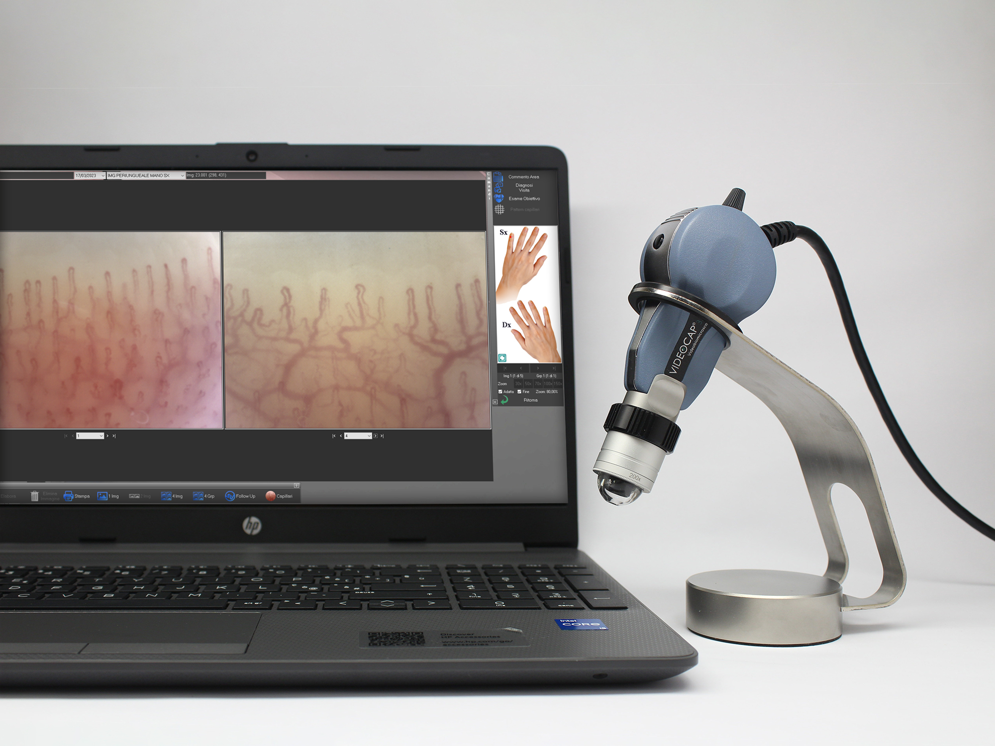
The Videocap® 4.0 D1 device is a video biomicroscope designed for epiluminescence, immersion, and polarized light imaging.
This portable, high-resolution videocapillaroscope is supplied with dedicated software that enables image acquisition and analysis of nailfold capillaries.
Its multidisciplinary versatility, comprehensive software, and portability make Videocap® 4.0 the ideal solution for professionals who require a flexible diagnostic tool for multiple applications.
The Kit
The Videocap® Reuma Basic Kit includes:
- Videocap® 4.0 D1 5MPX digital videocapillaroscope probe
- 200x lens complete with plexiglass dome
- Tabletop probe holder
- Latest-generation desktop or laptop computer
- Color laser printer
- Videocap® Reuma software
- Aluminum carrying case
- USB foot pedal for image capture
- Cedarwood oil for immersion, which reduces light dispersion and enhances image clarity and detail
- Photographic atlas of capillaroscopy
The Probe
The Videocap® 4.0 D1 is a video biomicroscope for epiluminescence, immersion, and polarized light applications.
It is the only device capable of performing advanced capillaroscopic analyses, thanks to its interchangeable lenses with various magnifications and the ability to switch between polarized and direct immersion light.
In rheumatology, this function is achieved using the 200x lens.
The optical system employs 12 LEDs to illuminate the examined area.
With the 200x optics, images are captured using cold white diode light through the immersion technique. Image quality is enhanced by applying cedarwood oil to the periungual area and by using the plexiglass dome, which ensures optimal contact and stability.
The optics feature a manual focus ring for precise adjustment.
For rheumatological use, optional fixed-magnification lenses of 100x or 300x, also equipped with plexiglass domes, are available.
Ergonomic, high-definition probe with excellent color fidelity and a lightweight design that reduces wrist fatigue during long working sessions.
The Videocap® 4.0 D1 connects to a PC via USB 3.0 and is powered directly from the USB port (4.5 – 5.5 VDC).
It includes a power on/off button located on the back of the camera.
Default brightness is set to automatic, but it can be manually adjusted using the dedicated dimmer knob.
- Camera sensor: 1/2.5” Rolling
- Resolution: 2592H × 1944V pixels (5 MPX – FULL HD)
- White balance
- Adjustable light intensity (rear dimmer control)
- Contrast
- Image gain
- Gamma
- Electronic image capture (via remote USB foot pedal)
The Videocap® 4.0 D1 complies with current medical device regulations:
Class I, Rule 10, Annex VIII – MDR 2017/745
Software
The Videocap® software allows image acquisition, storage, and archiving.
Developed to meet the needs of demanding professionals, it provides a complete tool for differential diagnosis and follow-up evaluation.
Its image optimization algorithm ensures high-quality visualization and photography with true color accuracy.
Secure and personal user access protected by password.
Register new visits, review archived ones, perform diagnoses, and print reports.
The software allows the comparison of images taken during previous visits.
Advanced search throughout the archive using keyword-based queries, categorized by gender, diagnosis, mapping, histological diagnosis, and clinical/capillaroscopic patterns.
User customization of body areas, diagnoses, account access, and all configuration settings.
Capture live images of the area under examination.
Acquired images can be edited by adjusting color, contrast, and brightness, or by applying graphic filters to enhance specific details.
Users can also add arrows, numbers, and text annotations directly to images for clearer interpretation.
The software supports import/export of images in .bmp, .jpeg, and DICOM formats.
Optional video recording module supporting AVI format on HDD or VHS.
Equipped with sophisticated measurement and image analysis tools, including:
- Measurement across multiple magnification ratios
- Capillary loop measurement
- Capillary density calculation within a user-defined range
Compare patient images captured during different visits, from different fingers, or imported from other sources (databases, CD-ROMs, user archives).
Generate complete and compliant reports in accordance with national and international guidelines.
Print 1-, 2-, or 4-image reports for the same lesion or follow-up examination.
All reports are fully customizable.
Compare previously acquired images with live images of the same area during follow-up sessions.
The Videocap® software can be installed in a network environment, allowing connection between multiple diagnostic workstations in multidisciplinary clinical settings.
