

Kit Videocap® Light Derma
Videocap® Light Derma
The Videocap® Light device is a video biomicroscope equipped with a polarizing filter and integrated optics.
Videocap® Light is a portable, high-resolution integrated system for videodermatoscopy — a simple, cost-effective solution that allows quick and accurate dermatoscopic examination and digital diagnosis of skin lesions.
The device is supplied with its dedicated software, which enables image acquisition of skin areas.
The Kit
The Videocap® Light Derma Kit includes:
- Videocap® Light digital videodermatoscope probe
- Integrated optics with 30x–60x–200x magnifications
- Spacers
- Videocap® Light Derma software
- USB foot pedal for image capture
- 5.0V/1A power supply and charger
- Carrying case
- Photographic atlas of dermatoscopy
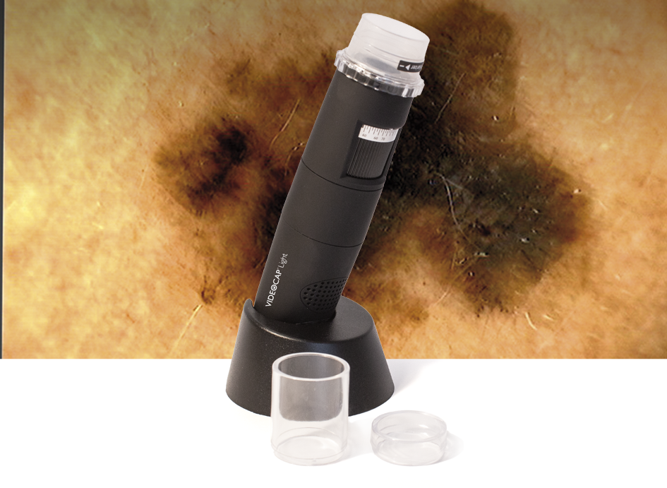
The Probe
Videocap® Light is a high-resolution video biomicroscope with an integrated polarizing filter.
The integrated lens provides 5x to 200x magnifications, making the device versatile and suitable for different areas of clinical investigation.
In dermatology, the optimal magnification for clear visualization is 30x.
The optical system uses 8 white LEDs with adjustable intensity to illuminate the area under examination.
Ergonomic, high-definition probe with a lightweight design for comfortable use and reduced wrist fatigue.
The Videocap® Light connects to a PC via USB 2.0 interface.
It is powered by a DC 5.0V/1A adapter, with a built-in lithium battery that provides approximately 2 hours of continuous use.
- Camera sensor: 1/2.5” CMOS
- Resolution: 5 MPX – 1280 × 1024 pixels
- White balance
- Gamma
- Exposure
- Light intensity adjustment
- Electronic image capture (remote foot pedal)
The Videocap® Light complies with current medical device regulations:
Class I – MDR 2017/745
Software
The Videocap® Light software enables image acquisition, storage, and archiving.
It has been developed to provide professionals with an advanced tool for differential diagnosis and follow-up evaluation.
Thanks to a dedicated image-saving algorithm, it ensures high-quality, color-accurate visualization both in live and captured mode.
Secure and personalized access protected by password.
Allows the registration of new visits, review of archived ones, and printing of visit reports.
Images from previous visits can be compared side by side.
User can customize account settings and configuration preferences.
Capture live images of the area under examination.
Images can be imported/exported in .bmp and .jpeg formats.
Compare patient images from different visits, body areas, or external sources (databases, CD-ROMs, user-created archives).
Generate complete and compliant reports according to national and international guidelines.
Reports can be customized with notes about body areas or diagnostic details.
The follow-up module allows comparison between a previously captured image and a live image of the same body area during a follow-up examination.