

Kit Videocap® Derma Phone
Videocap® Derma Phone
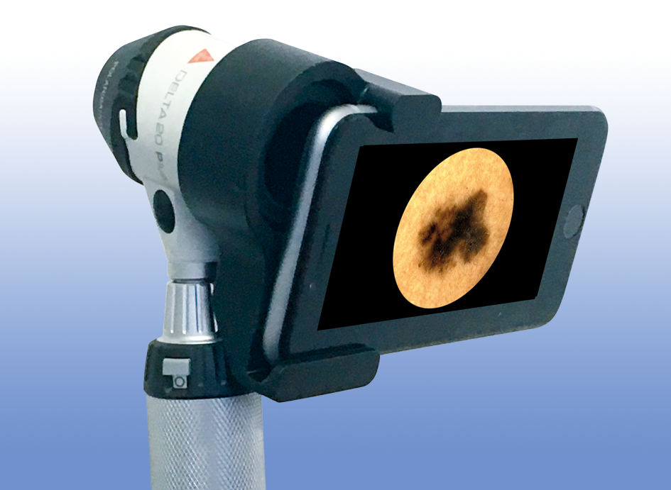
The Videocap® Phone configuration allows dermoscopic examination using the Heine Delta 30 integrated with a smartphone and the Videocap® Dermo Software.
This system provides everything needed for digital diagnosis of skin lesions with a simple click from your smartphone. It combines convenience and professionalism thanks to the integration with the Videocap software.
The Kit
The Videocap® Derma Phone Kit includes:
- Heine Delta 30 dermatoscope
- Heine Smartphone Adapter / Heine Delta 30
- Smartphone App
- Videocap® Derma Software
- >Wi-Fi Image Transmission Module (Phone)
- Latest-generation desktop or laptop computer
- Color laser printer
- Wi-Fi router
- Dermoscopy photographic atlas
The Dermatoscope
Heine Delta 30 is a high-precision manual dermatoscope using epiluminescence, ideal for detailed skin analysis.
With polarized light and optical magnification, it enables quick and reliable diagnosis — essential in dermatological prevention.
Thanks to the Videocap smartphone app, the dermatoscope can be connected to the dedicated software for image analysis and reporting.
Effective field of view of 30 mm, thanks to a 32 mm lens.
10x Magnification.
LED HQ illumination system with high color rendering, allowing operation at three different brightness levels, either in contact with or at a distance from the skin surface. The LED lighting ensures proper illumination and accurate color reproduction.
Ergonomic handle with an angled design. Compact and lightweight.
Switch from polarized to non-polarized light with a single button press — the “Toggle” function. Polarization ensures a glare-free view.
Rechargeable Li-Ion battery — no memory effect, charges easily regardless of charge level.
Includes a USB medical-grade power adapter.
The Software
The Videocap Software enables image acquisition, storage, and archiving.
It has been developed to provide even the most demanding professionals with a powerful diagnostic tool for differential diagnosis and follow-up evaluation.
Using a dedicated image storage algorithm, it guarantees high visual and photographic quality without color distortion.
User access is secure and personal, protected by a password.
Ability to record new visits, review those already stored in the archive, process diagnoses, and print visit reports. The software also allows comparison of images taken during previous visits.
The search section enables queries across the entire archive using a keyword-based system, with filters by gender, diagnosis, mapping, histological diagnosis, and clinical or dermoscopic patterns.
Users can customize body areas, diagnostic categories, account settings, and other interface options.
Capture live images of the area under examination.
Images can be saved and processed, adjusting color, contrast, and brightness or applying special graphic filters to highlight details.
Post-processing tools include annotations and geometric shapes (arrows, circles, rectangles, freehand selection, grid overlay).
The software supports dynamic localization and mapping on real images and body maps.
It also allows import/export of images in .bmp, .jpeg, and DICOM formats.
Optional video module enables recording in AVI format on HD or VHS.
Compare patient images from different visits, body areas, or even external sources (databases, CD-ROMs, personal archives).
Generate complete and accurate reports in line with national and international guidelines.
Reports can include 1, 2, or 4 images of the same lesion or visit, or follow-up documentation of a specific area.
All reports are fully customizable.
You can also print mole maps and dermoscopic images with corresponding references.
The follow-up module allows live comparison between a current image and one from a previous examination.
The Videocap Software can be installed on a network to meet the multidisciplinary needs of clinical facilities, allowing diagnostic workstations to be connected across departments.