

Kit Videocap® Derma Basic
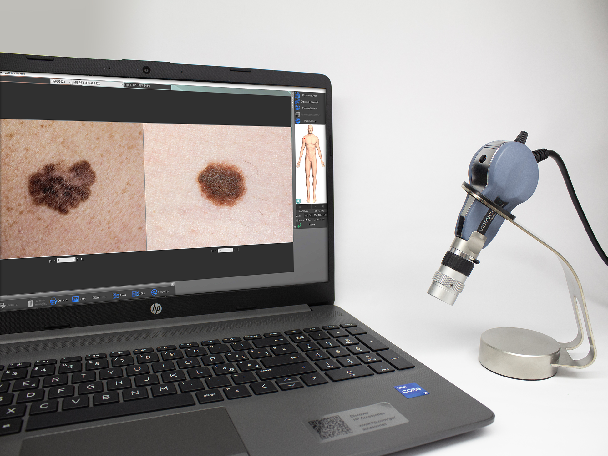
Videocap® 4.0 D1 Derma Basic
The Videocap® 4.0 D1 device is a video biomicroscope for epiluminescence, immersion, and polarized light imaging.
This portable, high-resolution videodermatoscope is supplied with dedicated software that allows image acquisition, storage, and analysis.
Its multidisciplinary versatility, comprehensive software, and portability make Videocap® 4.0 the ideal solution for professionals seeking a flexible diagnostic tool for multiple clinical applications.
The Kit
The Videocap® Derma Basic Kit includes:
- Videocap® 4.0 D1 5MPX digital videocapillaroscope probe
- 20x–50x polarized lens with 20x and 50x spacers (optional 30x spacer)
- Tabletop probe holder
- Latest-generation desktop or laptop computer
- Color laser printer
- Videocap® Derma software
- Aluminum carrying case
- USB foot pedal for image capture
- Photographic atlas of dermatoscopy
The Probe
The Videocap® 4.0 D1 is a video biomicroscope designed for epiluminescence, immersion, and polarized light imaging.
It is the only device capable of performing both dermatoscopic and trichoscopic analyses, thanks to interchangeable lenses with different magnifications and the ability to switch between polarized and direct immersion light.
In dermatology, this function is achieved using the 20x–50x optics, with specific spacers that allow observation of the area of interest at fixed magnifications of 20x and 50x (optionally 30x).
The optical system uses 12 LEDs for illumination of the area under examination. With the 20x–50x lens, images can be acquired using either white diode light or polarized light, by rotating the metal ring on the optics.
The optics feature a manual focus system operated via an adjustment ring.
Ergonomic, high-definition probe with excellent color fidelity, designed to reduce wrist fatigue during long working sessions.
The Videocap® 4.0 D1 connects to a PC via USB 3.0 and is powered directly from the USB port (4.5 – 5.5 VDC).
It features a power on/off button located at the back of the camera.
Default brightness is set to automatic, but it can be easily adjusted via the dedicated brightness control knob.
- Camera sensor: 1/2.5” Rolling
- Resolution: 2592H × 1944V pixels (5 MPX – FULL HD)
- White balance
- Adjustable light intensity (rear dimmer control)
- Contrast
- Image gain
- Gamma
- Electronic image capture (via remote USB foot pedal)
The Videocap® 4.0 D1 complies with current medical device regulations:
Class I, Rule 10, Annex VIII – MDR 2017/745
Software
The Videocap® software allows image acquisition, storage, and archiving.
It has been developed to provide professionals with an advanced tool for differential diagnosis and follow-up evaluation.
Its proprietary image-saving algorithm ensures high-quality visual and photographic results without color distortion.
Secure and personal access, protected by password.
Register new visits, review archived ones, perform diagnoses, and print reports.
The software allows comparison of images taken during previous visits.
Advanced search across the entire archive using keywords, grouped by category (gender, diagnosis, mapping, histological diagnosis, clinical and dermatoscopic patterns, etc.).
User customization of body areas, diagnoses, access accounts, and general configuration settings.
Capture live images of the area under examination.
Saved images can be processed by adjusting color, contrast, and brightness, or by applying graphic filters that highlight specific details.
Post-processing allows the insertion of annotations and geometric shapes (arrows, circles, rectangles, freehand selection, grid).
The software supports dynamic localization for mole mapping on real images and body maps.
Images can also be imported/exported in .bmp, .jpeg, and DICOM formats.
Optional module for AVI video recording on HDD or VHS.
Compare patient images taken during different visits, from different body areas, or from external sources (databases, CD-ROMs, historical archives).
Generate complete and accurate reports in accordance with national and international guidelines.
Print reports with 1, 2, or 4 images of the same lesion or visit, or follow-up comparisons.
All reports are fully customizable.
Possibility to print mole maps and dermatoscopic images with corresponding map references.
Compare a previously captured image with a live image of the same body area during a follow-up examination.
The Videocap® software can be installed in network environments, supporting multidisciplinary clinical settings and connecting multiple diagnostic workstations.