

Kit Videocap® Reuma Superior
Videocap®4.0 D1 Reuma Superior
The Videocap® 4.0 D1 device is a video biomicroscope designed for epiluminescence, immersion, and polarized light imaging.
This portable, high-resolution videocapillaroscope is supplied with dedicated software that allows the acquisition and analysis of nailfold capillary images.
Its multidisciplinary versatility, comprehensive software, and portability make Videocap® 4.0 the ideal solution for professionals who require a flexible tool for different diagnostic applications.
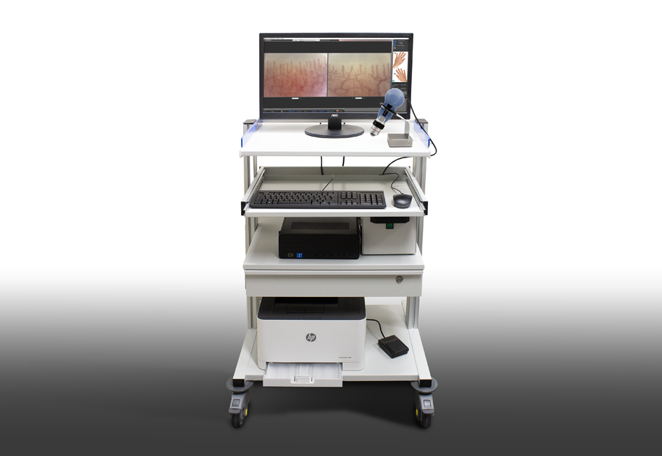
The Kit
The Videocap® Reuma Superior Kit includes:
- Videocap® 4.0 D1 5MPX digital videocapillaroscope probe
- 200x lens complete with plexiglass dome
- Latest-generation desktop or laptop computer
- Color laser printer
- Videocap® Reuma software
- Autocapi® module for automatic capillary counting
- Medical trolley with integrated workstation
- 1000W isolation transformer
- Aluminum carrying case
- USB foot pedal for image capture
- Cedarwood oil for immersion, which reduces light dispersion and enhances image clarity
- Photographic atlas of capillaroscopy
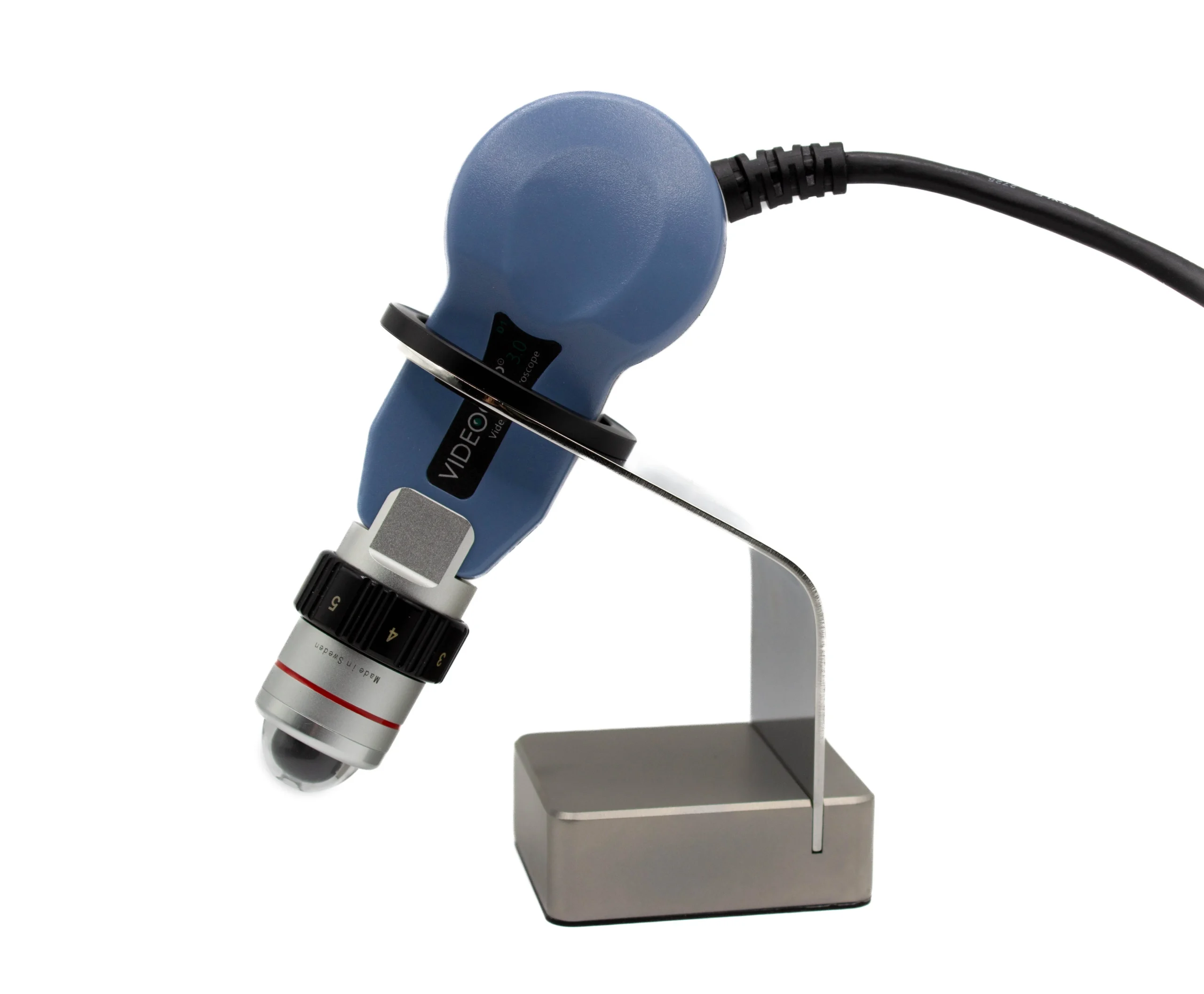
The Probe
Videocap® 4.0 D1 is a video biomicroscope for epiluminescence, immersion, and polarized light diagnostics.
It is the only device capable of performing advanced capillaroscopic analyses, thanks to its interchangeable lenses with various magnifications and the ability to switch between polarized and direct immersion light.
In rheumatology, the specific application is achieved using the 200x lens, optimized for nailfold microcirculation analysis.
The lens employs a 12-LED system to illuminate the area under observation. With the 200x optics, images are acquired using cold white diode light through the immersion technique. Image acquisition is enhanced by applying cedarwood oil to the periungual area and using the plexiglass dome for stabilization.
The optical system includes manual focusing via an adjustment ring.
In rheumatological studies, optional fixed magnification lenses of 100x and 300x, each equipped with a plexiglass dome, are also available.
Ergonomic, high-definition probe with superior color fidelity and lightweight design to reduce wrist fatigue during extended use.
The Videocap® 4.0 D1 connects to a PC via USB 3.0 and is powered directly through the USB port (4.5 – 5.5 VDC).
It includes a power on/off switch located on the back of the camera.
Default brightness is set to automatic, but can be easily adjusted using the dedicated control knob.
- Camera sensor: 1/2.5” Rolling
- Resolution: 2592H × 1944V pixels (5 MPX – FULL HD)
- White balance
- Adjustable light intensity (rear dimmer control)
- Contrast
- Image gain
- Gamma
- Electronic image capture (via remote USB foot pedal)
The Videocap® 4.0 D1 complies with current medical device regulations:
Class I, Rule 10, Annex VIII – MDR 2017/745
Software
The Videocap® software allows image acquisition, storage, and analysis.
Developed for demanding professionals, it provides an advanced tool for differential diagnosis and follow-up evaluation.
Its image-optimization algorithm guarantees high-quality visualization and photography without color distortion.
Secure, personal access protected by password.
Register new visits, review archived ones, perform diagnoses, and print reports.
The software allows comparison of images taken during previous visits.
Advanced search across the archive using keywords, grouped by categories (gender, diagnosis, mapping, histological diagnosis, clinical and capillaroscopic patterns, etc.).
User customization of body areas, diagnoses, accounts, and configuration settings.
Acquire live images of the examined area.
Captured images can be saved and enhanced by adjusting color, contrast, brightness, and applying graphic filters to highlight fine details.
Users can also annotate images by inserting arrows, numbers, and text labels for easier interpretation.
The software supports import/export of images in .bmp, .jpeg, and DICOM formats.
Optional module for AVI-format video recording to HDD or VHS.
Includes sophisticated measurement and analysis tools:
- Multi-scale magnification measurement
- Capillary loop size measurement
- Capillary density calculation within a selected area
Compare images of the same patient across different visits, from different fingers, or from external archives (databases, CD-ROMs, user-created archives).
Generate complete, standardized reports in accordance with national and international guidelines.
Print 1-, 2-, or 4-image reports for the same lesion or follow-up.
All reports are fully customizable.
Compare a previously saved image with a live image of the same area during a control visit.
The Videocap® software can be installed in a network environment, enabling multidisciplinary clinical setups and connecting multiple diagnostic workstations.
Autocapi® Module
Autocapi® is an optional software module for the Videocap® Rheumatology Suite, dedicated to automatic capillary counting.
It is included by default in the Videocap® Reuma Superior configuration.
Capillary density is a fundamental parameter for diagnosing vascular pathologies and evaluating treatment efficacy.
Until now, this assessment has been operator-dependent; however, Videocap® is the first software to feature automated capillary counting, providing objective and reproducible results.
This function is crucial for clinical research, as it enables standardized, quantitative analysis based on mathematical models and algorithms, removing subjective interpretation.
It also provides a reliable tool for therapy monitoring, allowing real-time verification of treatment effectiveness through accurate capillary quantification.
Clinical validation was achieved through the study:
“Automated assessment of absolute nailfold capillary number on videocapillaroscopic images: Proof of principle and validation in systemic sclerosis.”
