

Kit Videocap® Derma Mapping
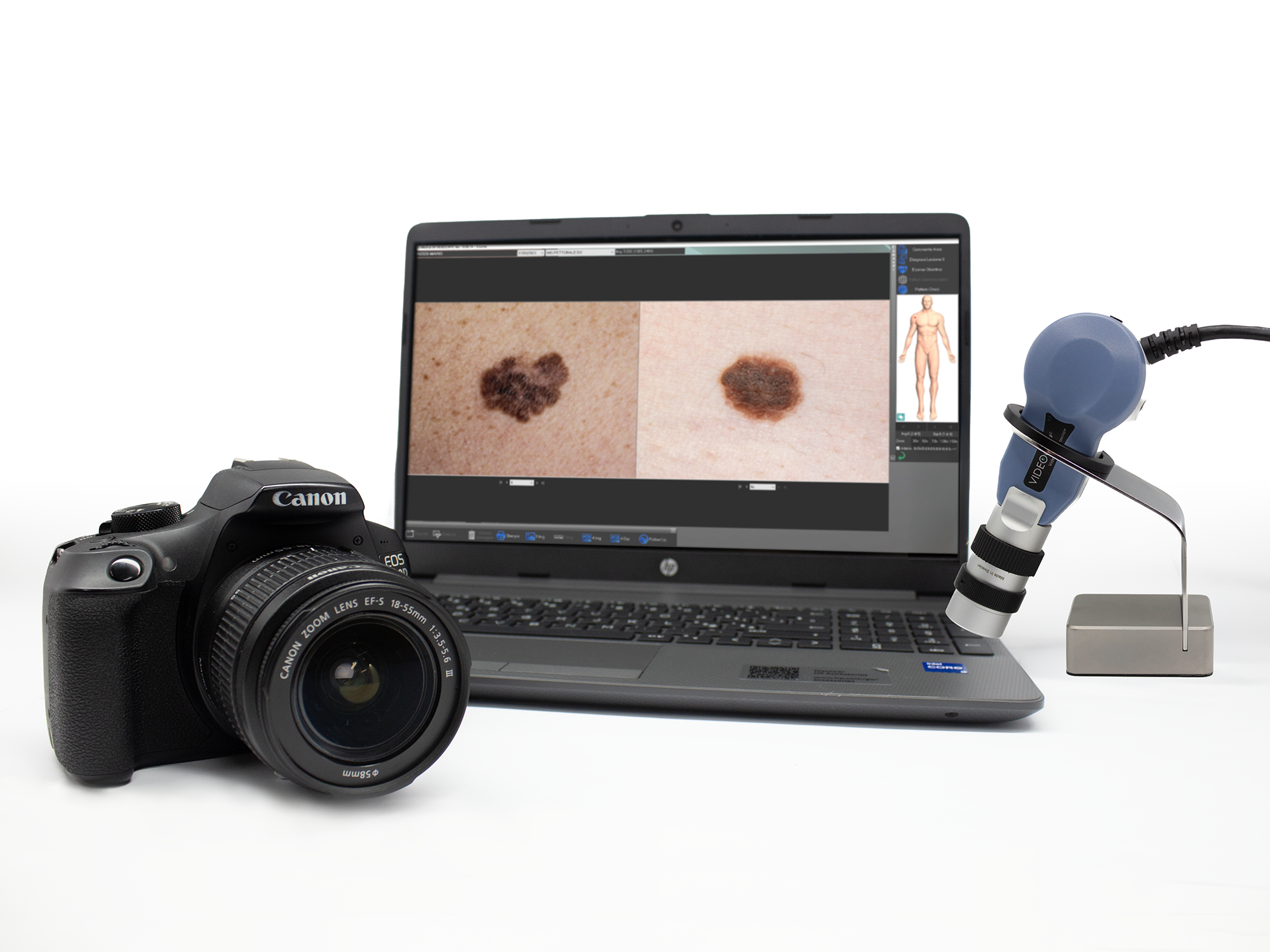
Videocap® 4.0 D1 Derma Mapping
The Videocap® 4.0 D1 device is a video biomicroscope for epiluminescence, immersion, and polarized light analysis.
This portable, high-resolution videodermatoscope comes with dedicated software that enables image acquisition, storage, and analysis.
In its “Derma Mapping” configuration, it allows the acquisition of dermatoscopic and macro images to perform mole mapping.
The Kit
The Videocap® Derma Mapping Kit includes:
- Videocap® 4.0 D1 5MPX digital videocapillaroscope probe
- 20x–50x polarized lens with 20x and 50x spacers (optional 30x spacer)
- >Canon DSLR camera (USB connection)
- Tabletop probe holder
- Latest-generation desktop or laptop computer
- Color laser printer
- Videocap® Derma software
- Aluminum carrying case
- USB foot pedal for image capture
- Photographic atlas of dermatoscopy
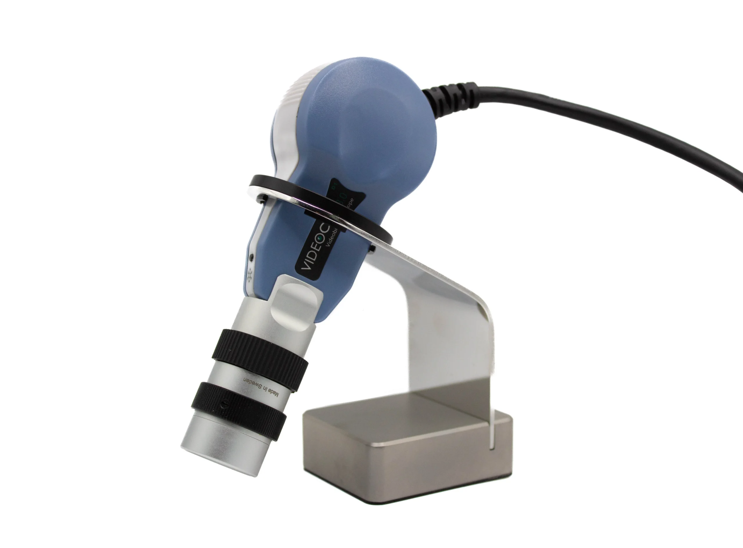
The Probe
The Videocap® 4.0 D1 is a video biomicroscope designed for epiluminescence, immersion, and polarized light diagnostics.
It is the only device capable of performing both dermatoscopic and trichoscopic analyses, thanks to interchangeable lenses of various magnifications and the ability to switch light types between polarized and direct illumination for immersion.
In dermatology, the device is used with the 20x–50x optics, featuring specific spacers that allow fixed magnifications of 20x and 50x (optionally 30x).
The lens uses a 12-LED illumination system for optimal lighting of the area under observation. With the 20x–50x lens, images can be captured under white diode light or polarized light using a metal ring on the optics.
Manual focus adjustment is available via the focusing ring.
Ergonomic, high-definition probe with excellent color fidelity and a lightweight design to reduce wrist fatigue during prolonged use.
The Videocap® 4.0 D1 connects to a PC via USB 3.0 interface and is powered directly through the USB port (4.5–5.5 VDC).
The probe features an on/off switch located at the back of the camera.
The probe brightness, set to “automatic” by default, can be easily adjusted using the dedicated dimmer knob.
- Camera sensor: 1/2.5” Rolling
- Resolution: 2592H × 1944V pixels (5 MPX) – FULL HD
- White balance
- Adjustable light intensity (rear dimmer control)
- Contrast
- Image gain
- Gamma
- Electronic image capture (remote foot pedal for image capture)
The Videocap® 4.0 D1 complies with current medical device regulations:
Class I, Rule 10, Annex VIII – MDR 2017/745
Software
The Videocap® software enables acquisition, storage, and archiving of images.
Developed to provide demanding professionals with an advanced tool for differential diagnosis and follow-up evaluation, it employs an optimized image-storage algorithm to ensure high-quality visualization and photography without color distortion.
Secure, personal user access protected by password.
Ability to register new visits, review archived ones, make diagnoses, and print reports.
Images from previous visits can be easily compared.
Comprehensive search system based on keywords, grouped by categories such as gender, diagnosis, mapping, histological diagnosis, clinical and dermatoscopic patterns, etc.
User customization of body areas, diagnoses, account settings, and usage configuration.
Live image capture of the area under examination.
Captured images can be saved and processed — adjusting color, contrast, and brightness, or applying graphic filters to highlight details.
Post-processing allows insertion of annotations and shapes (arrows, circles, rectangles, freehand selections, grid).
The software also provides dynamic localization for mapping on real images and bodymaps.
Images can be imported/exported in .bmp, .jpeg, and DICOM formats.
Optional video-recording module enabling AVI format recording on hard disk or VHS.
Compare patient images taken during different visits, from different body areas, or from external sources (databases, CD-ROMs, historical archives).
Generate complete reports according to national and international guidelines.
Print different types of reports — 1, 2, or 4 images of the same lesion or visit, or follow-up comparisons.
All reports can be customized.
Printouts may include the mole map and dermatoscopic images with their corresponding map references.
Allows comparison between a previously captured image and a live image of the same body area during a follow-up visit.
The Videocap® software can be installed in network environments, supporting multidisciplinary clinical settings by connecting multiple diagnostic workstations.